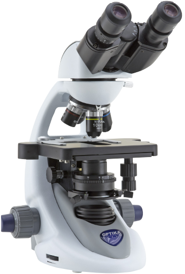
This series incorporates all the experience gathered by OPTIKA Microscopes in the field of light microscopy, adapted specifically for common laboratory applications. Suitable for routine microscopy with brightfield, darkfield, phase contrast and LED fluorescence, designed to last.
Special technology able to double the light intensity for incomparable performance, ensuring constant pure-white colour temperature (6,300K colour temperature). Relevant money and energy saving thanks to the incredibly low energy consumptions which allows you to cut the electricity bills by 90%. The electric consumption (3.6 W only) proves the high efficiency of this system incredibly high light intensity combined with low consumption.
Students and basic users will enjoy B-290 Series for the clear and sharp images they can get using 100x objective with water, thanks to the extremely bright X-LED3 light and the fully centerable Abbe condenser.
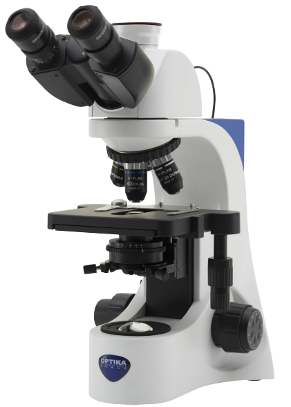
This series incorporates all the experience gathered by OPTIKA Microscopes in the field of light microscopy, adapted specifically for common laboratory applications. Suitable for routine microscopy with brightfield, darkfield (oil and dry), phase contrast, fluorescence and polarized light, designed to be extremely stable on the bench and last long.
OPTIKA B-380 phase contrast microscopes are equipped with a 5-position dedicated rotating condenser for brightfield (standard use), phase contrast (10x/20x, 40x and 100x phase diaphragms), and a darkfield position for dry objectives.
Achieve bright images, improved colour fidelity, pure white colour temperature, incredibly low energy consumptions and longer lifespan with the unique X-LED³ technology, able to double the light intensity for incomparable performance. Relevant money and energy saving thanks to the incredibly low energy consumptions which allows you to cut the electricity bills by 90%.
OPTIKA B-510 Series represents the ideal, ultra-modern, advanced routine microscope for efficient analysis in transmitted light applications, carefully engineered considering all the relevant aspects for users. This series offers user-friendly operations, robustness, durability and superb resolution, delivering contrasted and sharp images through the impressive IOS W-PLAN optics, the Infinity Corrected Optics (IOS) providing the best cost-effective choice for high contrast and resolution, matching all the requirements of labs requiring good quality routinary optics and designed to ensure field flatness up to F.N. 22.
The full Köhler system optimizes the microscope optical path to produce high sample contrast and homogeneous bright light, reducing image artifacts, whilst the state-of-the-art, exclusive X-LED3 lighting source makes sure your specimen will be properly and homogeneously illuminated with incredibly low consumptions and significantly long lifespan.
With multi-head observation systems, up to 5 people/colleagues can observe the same image on B-510; ideal for teaching and training students, especially in the medical field. The main observer and additional viewers will benefit of an extremely homogeneous light conditions, with a three-colour pointer with settable intensity to highlight points of interest.

B-810/B-1000 is built on IOS Infinity Corrected optical system, which gives both top-notch optical performances, and the possibility to extend your instrument with the broad range of accessories and modules. X-LED illumination is the best solution to have pure white light, very intense even at higher magnification, and optimum power efficiency given by solid state source. If you are a looking for our best solution to your present and future professional demands, B-810/B-1000 is the answer. Highest category of optical equipment among our product range guarantees a sharp and clear view in any situation, while top level mechanical design offers sturdiness and long lifetime. For different observation methods, the 5-position centrable rotating condenser for brightfield (standard use), phase contrast (10x/20x, 40x and 100x phase diaphragms), and a darkfield position for dry objectives represents a valuable solution, even for future upgradibility: one answer for multiple needs!
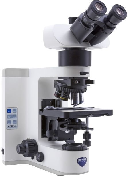
OPTIKA B-1000 Series is the result of an extensive modularity combined with the state-of-the-art, exclusive and high performing X-LED8 lighting source (8 W), corresponding to over 100 W halogen bulb.
This series features the outstanding brightness and incredible colour reproduction, to be a reliable solution for routine and research applications with up to a field of view of 24 mm. Fully modular, B-1000 Series gives multiple options of configuration, from basic manual controls to a fully motorized version, including motorized nosepiece, stage and focus. Its capabilities go further, with the possibility to install the exclusive Automatic Light Control (ALC) for unparalleled performance.
The selection of nosepieces, objectives, stages and condensers is extremely wide, offering several options to create the most suitable microscope for every need! B-1000 is also configurable as multi-head teaching discussion microscope, providing multiple observations up to 10 heads simultaneously. A perfect instrument for teaching, offering the main head complete of an LED movable pointer (RGB) to simplify the work of the main observer when needs to highlight and show some specific aspects of the specimen.
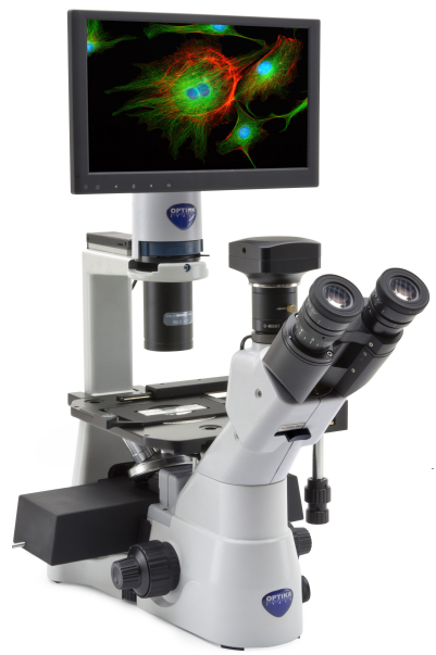
Inverted microscopes are useful for observing living cells or organisms at the bottom of a large container (e.g., a tissue culture flask) under more natural conditions than on a glass slide, as is the case with a conventional microscope. IM-3 Series includes a version for simultaneously brightfield and phase contrast method, engineered and designed to be your ideal solution for fast and reliable routine inspections.The glass stage surface allows an optimal visual access to the objective turret. A particularly simple and ingenious optical design allows stable alignments and smooth and accurate movements.
X-LED8 illumination system is based on a pure white high-efficiency LED and a special optics. It guarantees constant color temperature (6300K) for all intensity levels, no heat generation, and extreme electrical comsumption efficiency. The whole system is pre-aligned and has a lifetime of 50,000 hours. The F.O.V. (field of view) of IM-3 Series is based on a very comfortable diameter of 22 mm. A natural and easy view is ensured, especially when typically required in a laboratory environment.
.jpg.jpg)
OPTIKA IM-5 is a new inverted research microscope producing brilliant images for the examination of living cells, organisms and several other specimens in large flasks, combining brightfield, darkfield and phase contrast techniques for the most demanding users.This inverted research microscope drives to new horizons providing Köhler condenser, ergonomic handy controls and significant unique features, such as the highest F.O.V. available on an inverted microscope (F.N. 24 mm). IM-5 is freely configurable in terms of objectives, by choosing among:
– IOS LWD W-PLAN (Plan-Achromatic LWD for brightfield, F.N. 22)
– IOS LWD W-PLAN PH (Plan-Achromatic LWD for phase contrast, F.N. 22)
– IOS LWD U-PLAN F (Semi-Apochromatic LWD for brightfield, F.N. 25)
– IOS LWD U-PLAN F PH (Semi-Apochromatic LWD for phase contrast, F.N. 25)

This model was created to meet all the needs related to research in life science and designed to be complemented by a series of packages dedicated to more advanced individual applications. For all intents and purposes, IM-7 is to be considered as an inverted imaging platform, due to its high expandability and state-of-the-art quality. The six-position nosepiece has a slot (in each of the six positions) for inserting DIC prisms. The filter turret can hold up to six fluorescence filterblocks. It is easily extractable, in order to facilitate the operation of inserting or replacing the filterblocks. The wide 3-layer mechanical stage comes with several interchangeable plates for the use of Petri dishes, flasks and slides. The movement of the stage is controlled by a long tilting handle equipped with a pair of knobs for X/Y axes. The universal condenser is a 6-position type, designed for brightfield, phase contrast and DIC.

The polarized light microscope is designed to observe specimens that are visible primarily due to their optical anisotropic or birefringent features. Polarized light microscopy is perhaps best known for its applications in the geological sciences, which focusses primarily on the study of minerals in rock thin sections. However, a wide variety of other materials can be examined in polarized light, including both natural and industrial minerals, concrete, ceramics, mineral fibers, polymers, starch, wood, urea. The technique can be used both qualitatively and quantitatively with success, and is an outstanding tool for the materials sciences, geology, chemistry, biology, metallurgy, and even diagnostic (gout analysis). Polychromatic light being viewed at the analyser, one specific wavelength will have been removed. This information can be used in a number of ways:
– If the birefringence is known, then the thickness, t, of the sample can be determined
– If the thickness is known, then the birefringence of the sample can be determined
As the order of the optical path difference increases, then it is more likely that more wavelengths of light will be removed from the spectrum. This results in the appearance of the colour being “washed out”, and it becomes more difficult to determine the properties of the sample. This, however, only occurs when the sample is relatively thick when compared to the wavelength of light.
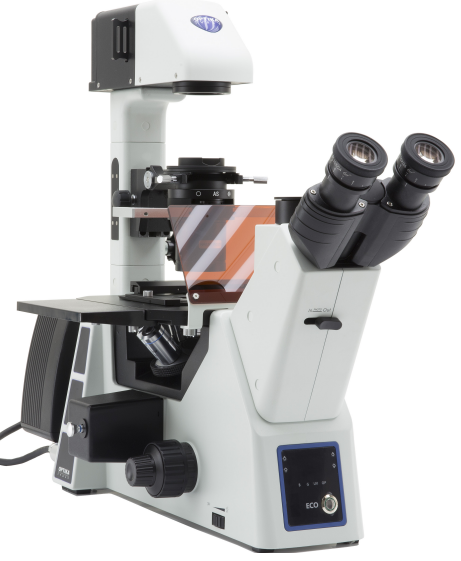
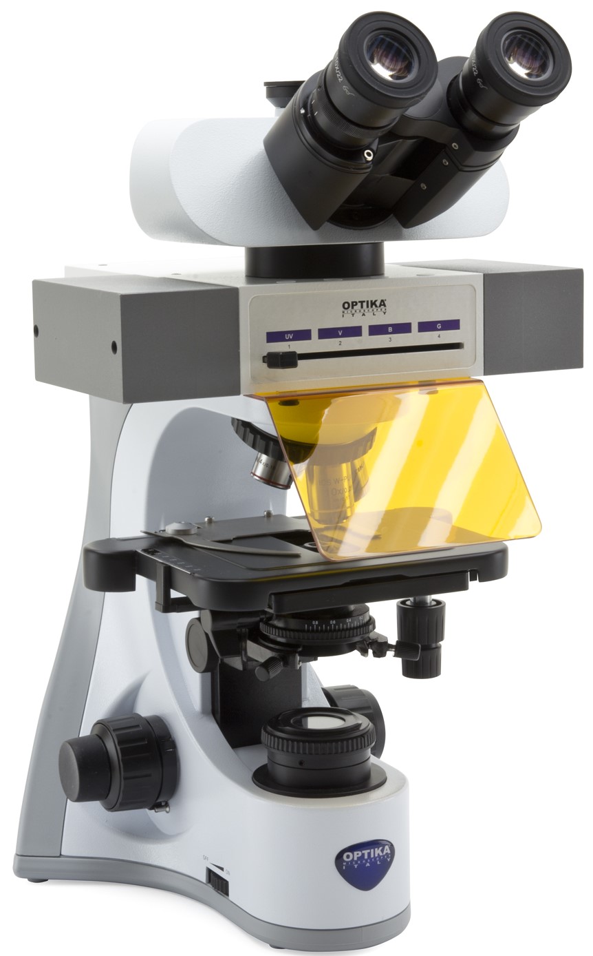
A fluorescence microscope is an optical microscope that uses fluorescence and phosphorescence instead of, or in addition to, reflection and absorption to study properties of organic or inorganic substances. The “fluorescence microscope” refers to any microscope that uses fluorescence to generate an image. The Epi Fluorescence microscope is equipped with a fluorescence illuminator wich generates incident fluorescence light.
Typical components of a fluorescence microscope are a light source (HBO mercury-vapor lamps are common; more advanced forms are high-power LEDs), the excitation filter, the dichroic mirror, and the emission filter. The filters and the dichroic mirror are chosen to match the spectral excitation and emission characteristics of the fluorophore used to label the specimen. In this manner, the distribution of a single fluorophore (color) is imaged at a time. Multi-color images of several types of fluorophores must be composed by combining several single-color images. Most fluorescence microscopes in use are epifluorescence microscopes, where excitation of the fluorophore and detection of the fluorescence are done through the same light path (through the objective). These microscopes are widely used in biology and are the basis for more advanced microscope designs
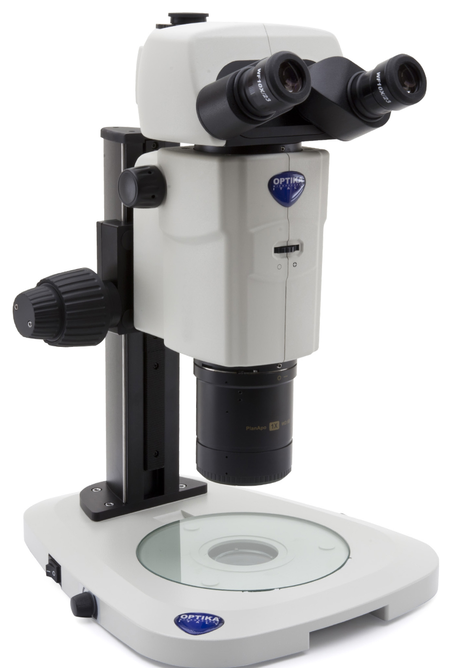
The SZR-180 is the top-of-the-range stereo zoom microscope dedicated to the world of research.
With a wide magnification range and high optical resolution combined with effective ergonomics, the SZR-180 is the perfect companion in advanced research. The SZR-180 with its 18:1 zoom ratio puts it at the top of its class. The wide range of magnifications available (7.5x-135x), coupled with a precise click-stop mechanism for working with reproducible zoom settings, makes it ideal for both low-magnification observation and high-magnification screening and observation of small cell structures. The Plan APO 1x apochromatic main objective provides high-resolution images at both low and high magnifications, making it ideal for observing and capturing images of the smallest details free of aberrations and halos. Combined with the zoom and the generous 23mm field number eyepieces, this provides a large FOV even at high magnifications (1.7mm).